We apologize for delays in customer service and transactions.
We are updating our hospital's information systems, and this may temporarily cause delays in customer service and transactions. We apologize...
Read moreMon–Fri 8–16

Magnetic resonance imaging (MRI) is the best method for determining the local spreading of prostate cancer. At Docrates Cancer Center, a new method of robot-assisted MRI-guided biopsy allows taking biopsies directly from abnormal lesions.
In the past decade, prostate MRI has developed vastly due to multiple‑parameter techniques, such as diffusion imaging, dynamic angiography and spectroscopy. Combined with these techniques, MRI has been ascertained to be the best currently available prostate cancer imaging method. MRI is the most effective imaging method for detecting tumors in the peripheral zones of the prostate, in particular.
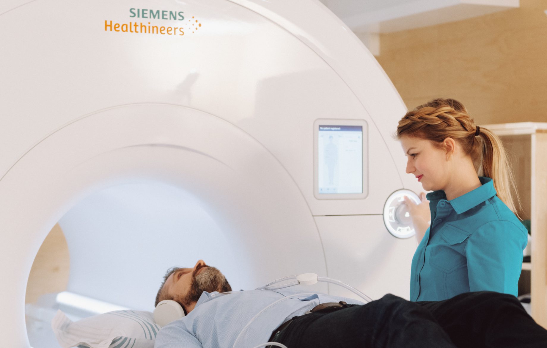
At Docrates, MRI scans are performed with next-generation MRI scanners. We use the Siemens Magnetom Vida 3T with 3-tesla imaging power. The 3 teslas provide more accuracy in the diagnosis of breast cancer and prostate cancer, in particular. The better images of the new MRI scanner enable physicians to make a quick and more detailed diagnosis without the need for further examinations. This also helps us draw up even better treatment plans.
In order to treat prostate cancer effectively, it is important to get a reliable picture of the spreading of the cancer before creating a treatment plan. A particularly important aspect of this is to study the relationship between the tumor and the prostatic capsule. An MRI scan shows whether the cancer has spread beyond the prostate. Therefore, an MRI scan is always required before making the final decision on prostatectomy surgery or radiotherapy. In addition, MRI scans can be useful at an early stage where the presence of cancer needs to be determined or before taking a biopsy.
An MRI scan is always performed in the planning phase of targeted radiotherapy. If an MRI scan shows that the cancer has spread to the seminal glands, this must be take into account in radiotherapy by targeting a larger dose of radiation to the glands. Whenever any risk factors concerning the recurrence of cancer, such as local spreading, are detected, hormonal therapy is always a valid option.
In order to make a final diagnosis and to determine the degree of malignancy, a pathologist must always study biopsies of any abnormal lesions.
A conventional set of biopsies includes approximately 12 biopsies from random sections of the prostate. This includes a risk of complications and infections. Patients typically experience pain and bleeding after the biopsy. At Docrates Cancer Center, a new method of robot-assisted MRI-guided biopsy allows taking biopsies directly from abnormal lesions, reducing the number of required biopsies to 2 or 3. The method is more accurate than fusion biopsy. Robot-assisted MRI-guided prostate biopsy is performed under local anaesthesia. No pain is inflicted on the patient.
We are updating our hospital's information systems, and this may temporarily cause delays in customer service and transactions. We apologize...
Read more
Docrates Cancer Center will open its first clinic in Sweden. Located in Stockholm, the new clinic will offer Swedish patients...

Reconstructing a removed breast is part of the treatment path of many breast cancer patients. The goal of the surgery...

New forms of treatment based on molecular diagnostics enable more and more patients to completely recover from cancer, and make...
Contact us!
Mon-Fri 8:00–16:00
