The number of Norwegian patients increased in 2023
Docrates Cancer Center has been treating Finnish and international patients in Helsinki for more than 15 years. In 2023, the...
Read moreMon-Thu 8-18, Fri 8-16

A tumor which has started to develop in the brain, is called a primary brain tumor. About half of these primary brain tumors belong to the group of glial or supportive tissue tumors called gliomas.
About 2/5ths (40%) of gliomas are glioblastoma, previously called glio¬blastoma multiforme. In the new 2021 WHO classification of brain tumors an astrocytoma grade (= class) IV is also an entity. In Finland, around 1000 brain tumors are detected annually, in Sweden about 1400. This means that approximately 150-200 glioblastomas are detected per year in Finland. Almost all glioblastomas occur in older adults. The average age to get a glioblastoma is about 60 years. About 10% of glioblastomas originate from other gliomas, such as astrocytoma, which over time become more malignant; these patients are usually younger, on average 45 years old.
Glioblastomas can grow in all parts of the brain, usually just below the cerebral cortex (=outer layer of the brain) in the white substance. Glioblastomas are most commonly found in the temporal, parietal or frontal lobe; in adults rarely in the brainstem or cerebellum.
It is not known why glioblastoma starts to grow. The only known risk factors are previous radiation therapy to the brain and some very rare genetic disorders. It is also not known from which cells glioblastoma begins to grow. It has been suggested that the origin could be so-called stem cells or some other more primitive precursor cells where a DNA or genetic damage has occurred, which the body has not been able to repair. Nowadays it is known that many cells in the brain can go into the so called stem cell -stage. The picture has consequently become more complicated. Glioblastoma cells are very variable, no cells are typical of glioblastoma.
In magnetic resonance imaging (MRI), glioblastomas are usually diffuse and irregularly shaped and non-uniform. They are often bulky. They often have a contrast-enhanced, circular area around the necrotic area, as shown. They often extend widely and may be surrounded by extensive water containing, swollen brain tissue. In many cases, the image is typical, but not always. Metastases of other cancers in the brain area and sometimes even meningeoma tumors of the meninges may look similar. Based on previous studies, it has been estimated that approximately 10% of tumor types will be misestimated based on imaging. Thus, diagnosis cannot be made reliably solely on the basis of imaging alone.
Glioblastomas are often large, heterogeneous tumors. Usually they have no clear boundaries. They often contain new blood vessels, but also areas that have gone into necrosis ie. died. When looking under a microscope, the tumor cells are very variable. In addition to small areas of necrosis, there are new, deformed blood vessels, many kinds of nuclei, pseudo-cellular, interconnected cells and cells that are dividing. In other glioblastomas the cell image can be more uniform. The former name glioblastoma multiforme describes the pathological picture. Multiforme means many forms. The margin to normal brain is not precise.
There areusually tumor cells in the area around the main tumor. This means that there usually are single malignant tumors in the brain tissue around the main tumor. In MRI images this brain tissue may look normal.
Although a glioblastoma can be suspected on the basis of the MRI scan, the definite diagnosis requires a sample of tissue that the pathologist evaluates. The pathologist describes the tumor’s microscopic appearance as well as the number of cells that divide, ie. malignancy. In addition, chromosomes and certain gene defects are determined from the tumor.
The degree of malignancy of glioblastoma is IV (four) the most malignant of gliomas. Approxi¬mately 10% of glioblastomas have such gene defects that they are considered to have developed of class II or III tumors. These, so-called secondary glioblastomas, generally have IDH1 or 2 mutations, a gene defect. Their prognosis is better than that of the non-mutated tumors. These IDH1 and 2 enzymes are important for cellular energy conversion. We are just beginning to understand the significance of this phenomenon and to exploit it in treatments. Another enzyme of great importance is MGMT (methylguanylmethyltransferase). MGMT’s role is to repair cell damage caused by, for example, radiation therapy or chemotherapy. If MGMT is methylated (i.e the regulating gene is methylated), MGMT cannot function. MGMT methylated tumors are therefore more sus¬ceptible to chemotherapy and the prognosis is better.
The pathologist can also evaluate how many of the tumor cells appear to divide. This is done by evaluating the Ki-67 percent. This percentage tells which proportion of the cells show K1-67 protein on cell surface which tells that they are dividing. Ki-67 protein does not occur on surface of resting cells. Usually, Ki-67 is high in glioblastoma, 20-30% or higher, but this percentage may and usually varies in different parts of the tumor.
Glioblastoma may have been growing for years or just a few months before it is diagnosed. The most common symptoms are various kind of seizures, paralysis symptoms, and disturbances in the executive functions such as judgment, exclusion of unnecessary stimuli, and tactfulness or flexibility. Seizures can often be very short, up to 1-2 minutes, and focal, such as a strange sense of smell or, for example, numbness of upper limb. Sometimes, on the other hand, seizures are generalized, ie in addition to jerky movements, there is a drop in the level of consciousness. Sometimes patients have a new type of headache. Headache is, however, very rarely the only symptom. Symptoms vary depending on the location and size of the tumor. For more details, see Brain Tumor Symptoms, click
If the tumor is located so that it can be operated, this is usually done. Normally, as much as possible is removed from the tumor without causing too many symptoms. If the tumor is located in a very difficult to operate place or the patient is in poor condition, only a sample can be taken.
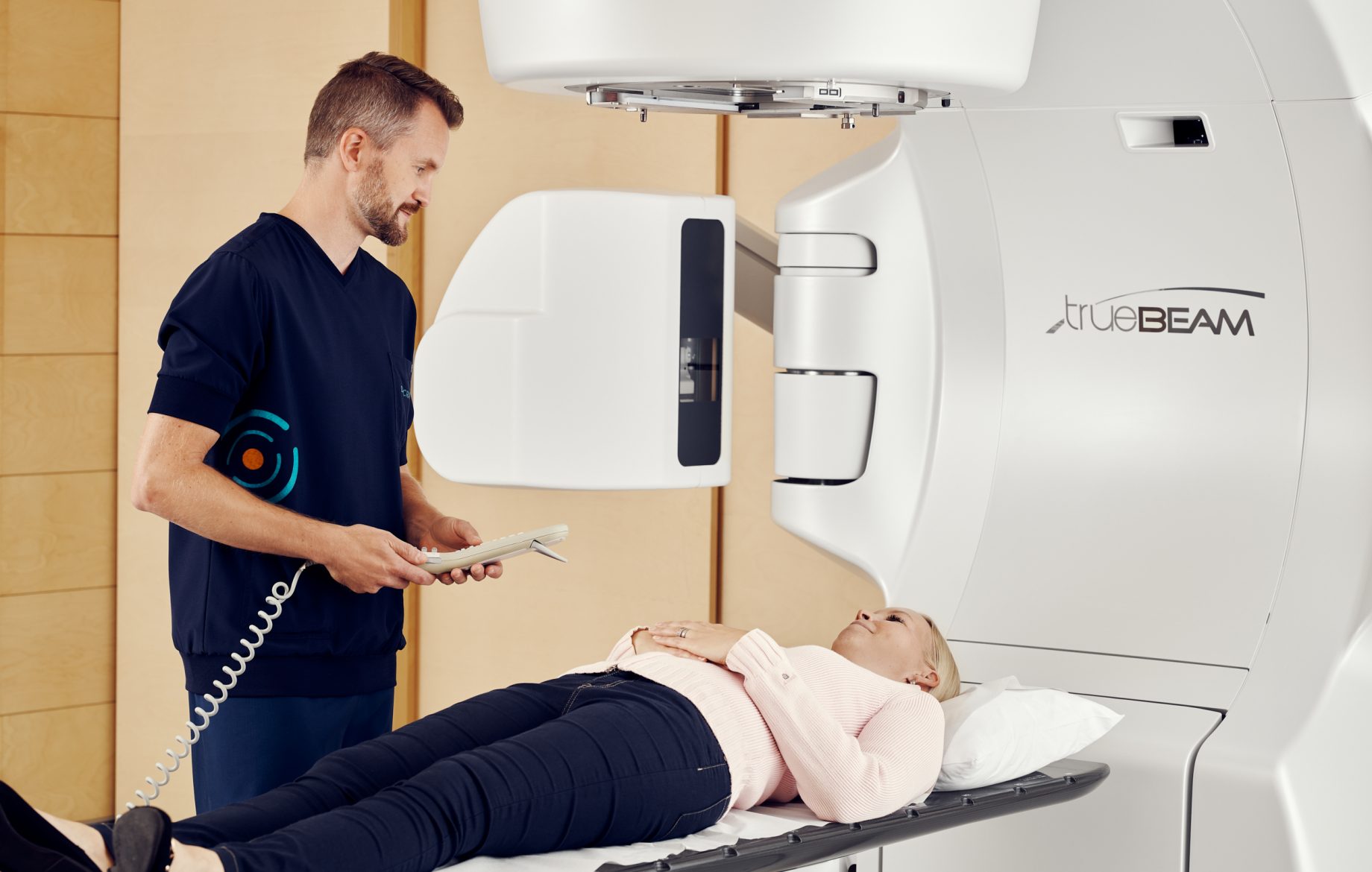
The prognosis for glioblastomas was substantially improved when radiotherapy was started in combination with chemotherapy. This so-called chemoradiation was developed in the early 2000s. In particular, this treatment has increased the number of patients living more than 2 or 3 years. If the patient is older and / or in poorer condition, shorter radiotherapy is often used and in some cases only chemotherapy is given, especially if the tumor has a MGMT mutation, i.e. it is more sensitive to chemotherapy. The most commonly used chemotherapeutic agent is temozolamide, which is administered orally on an empty stomach and is penetrates well into the brain.
Treatment strategies for recurrent tumors vary widely. Reoperation is offered to patients in good condition, additional radiotherapy can often be given, or chemotherapy restarted. In addition, there are experimental treatments.
Docrates Cancer Center has been treating Finnish and international patients in Helsinki for more than 15 years. In 2023, the...
Read more
Docrates Cancer Center joined forces with Medanta, a pioneer in healthcare workwear, to create a more functional workwear collection for...

Riikka Talsi, M.Sc. (Tech), who defended her doctoral thesis at Aalto University School of Science, Department of Industrial Engineering and...

Cancer. The word alone makes the heart beat faster and fills people's minds with questions. What if I have cancer?...
Contact us!
Mon-Thu 8:00-18:00, Fri 8:00-16:00
