The examination always starts with low-dose computed tomography (CT), which is followed by a PET imaging stage. The tracer injection is given intravenously, after which PET imaging is carried out. The characteristics of cancer tissue deviate from normal tissue, and PET imaging allows indicating the metabolic differences of cancer tissue as pictorial information.
In the case of specific cancers, the PET-CT imaging may be performed not only in an ordinary fashion, but also paced with breathing, in which case as precise an image as possible is obtained of the object by computing the inaccuracy caused by breathing away from the images. The images are fused, i.e. the computer combines the CT and PET images.
The total duration of the PET-CT examinations is approximately two to three hours, depending on the type of examination. After administering the tracer, you must wait for the tracer to be taken up by its target via metabolism. Most usually, body imaging as a whole takes about 25 minutes.
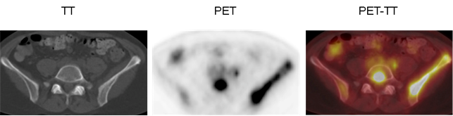
In the combined PET-CT image, the tracers clearly shine and indicate where the cancer tumors are located.