


The number of Norwegian patients increased in 2023
Docrates Cancer Center has been treating Finnish and international patients in Helsinki for more than 15 years. In 2023, the...
Read moreMon-Thu 8-18, Fri 8-16
Computed tomography (CT) is one of the most important imaging methods in diagnosing cancer, investigating tumor staging and following up on cancer treatment. A CT scan can be used as a primary or a supplementary examination. CT scans can also be used to examine other disorders and illnesses.
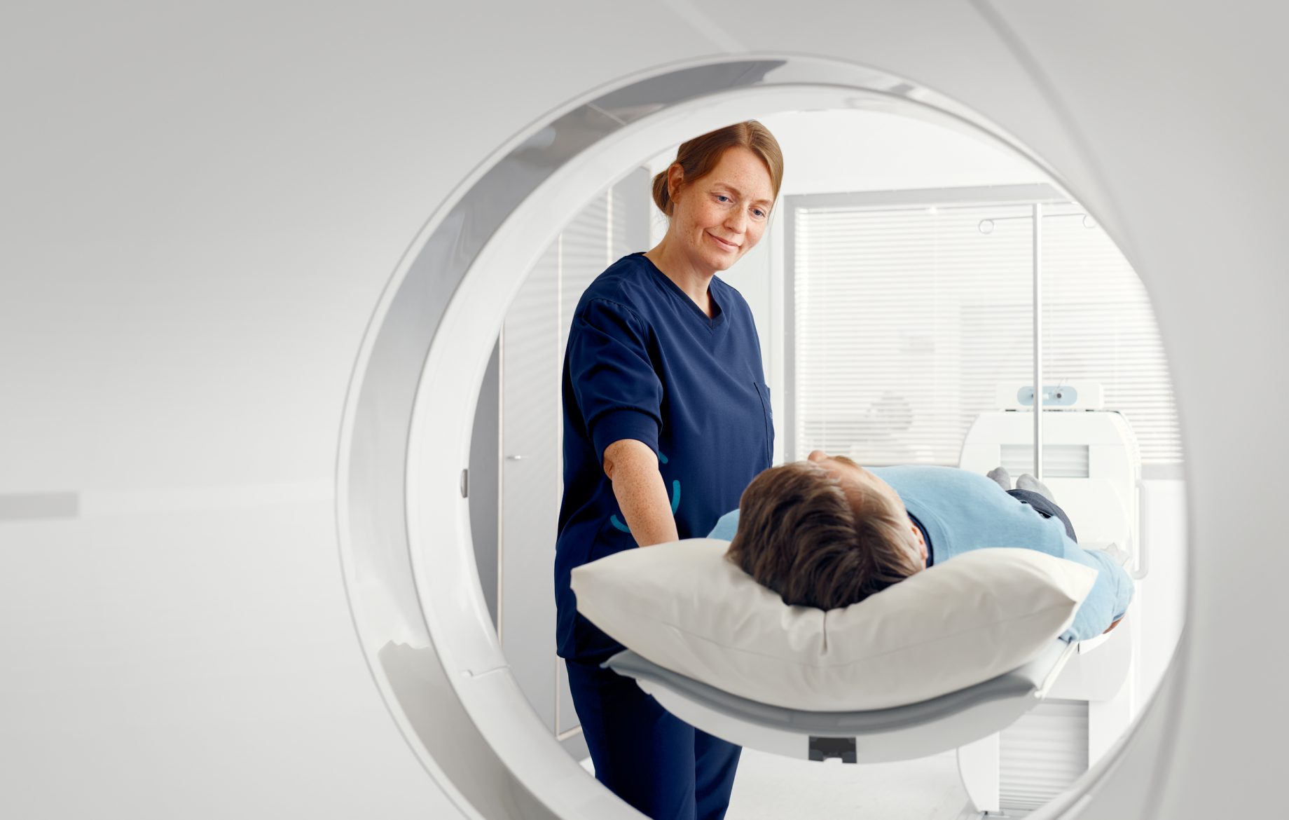
During the scan, the patient lies on a table that slides into and through the scanner. The rotating part in the CT Scanner includes an x-ray tube that emits the x-rays used for the scan. On the opposite side of the scanner, a sensor measures the radiation transmitted through the patient. The rotating time of the scanner part can be adjusted as needed, ranging from about 0.25 to 4 seconds. As the x-rays transmitted through the patient are measured from several angles during the rotation, the data can be compiled into a cross-sectional image to detect any features suppressing the radiation through the tissue as so-called Hounsfield units (HU). In CT scans, air usually appears black (HU=-1000), water as medium grey (HU=0) and bone or metal as clear (HU > 1000). The greyscale of the image, possibly containing more than 65,000 shades, helps examine details in both the light and the dark areas.
The imaging table usually moves while the radiation is produced and the transmission is measured. This is called a spiral scan. Known as a particularly quick imaging option, a spiral scan is the most common method used in CT scans. Another option is to scan the cross-sectional images or slices by keeping the table still during radiation and measuring. Once the tube and the sensor have completed a full circle, the table is moved and a new section is scanned.
While the CT machine primarily produces cross-sectional images, the large amount of data collected in the examination makes it possible to compute the slices in any direction. The thickness of the slices can also be adjusted after the scan. The images can also be used to create three-dimensional models, if necessary.
A CT scan is a quick process; one scan takes about 10–60 seconds. The patient’s entire body can be scanned while they hold their breath. A CT scan can include one or multiple positioning scans and, usually, 1–3 actual scans. The preparations for the scan usually take more time than the actual scan.
In August 2020, we acquired and installed a cutting-edge Siemens CT scanner as part of our new PET-CT equipment. The CT equipment in the combined scanner is a diagnostic, 128-slice device similar to the Somatom Definition Edge by Siemens.
The new equipment enables shorter scan times, smaller radiation doses and more detailed images, which translates into improved patient safety and comfort as well as more precise diagnostics. The new image computation methods make it possible to reduce both the radiation dose and the noise in the image. This allows us to better optimise the image quality and the radiation dose. Distortion caused by metal objects in the body (hip prostheses, implants, screws) can often be completely eliminated or its impact can be reduced without affecting diagnostic accuracy.
Docrates Cancer Center also uses a Siemens SOMATOM Sensation, a 24-slice CT scanner particularly efficient for dose planning in radiotherapy. The Sensation also enables all types of usual diagnostic scans. The large scanner bore, 82 cm in diameter, allows optimal positioning for radiotherapy-related imaging.
Docrates Cancer Center provides you prompt and easy access to imaging scans with a referral from a specialist. We provide high-quality, individual diagnostics and latest cancer treatment without delay. Docrates Cancer Center specializes in cancer imaging.
Most CT scans use an iodinated contrast agent. The agent efficiently suppresses radiation, transforming the contrast between tissue so that the examined part stands out. The health of the kidneys and any allergies to the contrast agent must be investigated before performing a CT scan with contrast agent. If the kidneys’ ability to filter blood is significantly impaired, the CT scan can be performed without a contrast agent.
The contrast agent is usually injected into a vein using an automated injector. The timing of the injection in relation to the start of the CT scan depends on the examination. Usually, the contrast agent is injected a few tens of seconds before the start of the CT scan, but the time may vary from a couple of seconds to several minutes. In some examinations, the patient drinks the contrast agent so that it is delivered into the stomach and the intestines instead of the veins.
A CT scan can only be performed with a referral from a specialist. Both the doctor providing the referral and the radiologist in charge of the scan will assess the risks of the scan in proportion to the benefits. The scan can only be performed if the assessed benefits outweigh the risks.
The scan is performed by a radiographer according to the instructions of the radiologist in charge.
The scan itself is painless and the patient does not feel anything abnormal. When using a contrast agent, the patient may notice a sense of warmth where the agent was injected or throughout their body. Some patients may also have a metallic taste in their mouth. These sensations usually pass quickly.
On some rare occasions, the iodinated contrast agent may cause an allergic reaction. The severity of the reaction may vary from mild to severe symptoms. If the patient has previously experienced a sudden reaction to an iodinated contrast agent, the CT scan can be performed without a contrast agent. The imaging department always carries the necessary medication, several radiographers and a doctor available in case any symptoms arise.
A CT scan is based on the use of x-rays, and the radiation dose of a CT scan is larger than the dose of a traditional x-ray. The typical effective dose from CT scanning the body is about 8 mSv, corresponding to about two and a half years of background radiation. One CT scan of the body for a 30-year-old patient increases the existing, lifelong cancer risk by about 0.09%, for a 50-year-old by about 0.05% and for a 70-year-old by about 0.03%. If a person had ten CT scans every year throughout their adult life (ages 18–65), the impact on their life expectancy would be smaller than that of being 25% overweight.
CT scans, like any examinations, may reveal changes that turn out to be benign in follow-up tests. These are called false positives. A false positive may cause concern and unnecessary follow-up tests, but it is also important to carry out the follow-up tests thoroughly.
Docrates Cancer Center has been treating Finnish and international patients in Helsinki for more than 15 years. In 2023, the...
Read more
Docrates Cancer Center joined forces with Medanta, a pioneer in healthcare workwear, to create a more functional workwear collection for...

Riikka Talsi, M.Sc. (Tech), who defended her doctoral thesis at Aalto University School of Science, Department of Industrial Engineering and...

Cancer. The word alone makes the heart beat faster and fills people's minds with questions. What if I have cancer?...
Contact us!
Mon-Thu 8:00-18:00, Fri 8:00-16:00