The temporary technical problem in the data network connections
A temporary technical problem occurred in the data network connections of Docrates Cancer Center. We apologise for any delays caused...
Read moreMon–Fri 8–16

A breast MRI is one of the most sensitive methods for diagnosing breast cancer. It can reveal cancer not seen on mammography.
A breast MRI is one of the most sensitive methods for diagnosing breast cancer. An MRI can be conducted as a primary examination, even before taking a needle aspirate sample, but it can also be conducted after the sample.
A breast MRI provides more information on the size and tumor staging of the cancer. An MRI can also detect cancerous growth not found in mammography, either in the same breast as the diagnosed cancer or in the other breast. In addition, an MRI may provide valuable information to the breast cancer surgeon when planning a so-called oncoplastic breast-conserving surgery. In the situations where the treatment starts with chemotherapy (neoadjuvant chemotherapy), the response to treatment is monitored with MRI scans preceding and during the treatment.
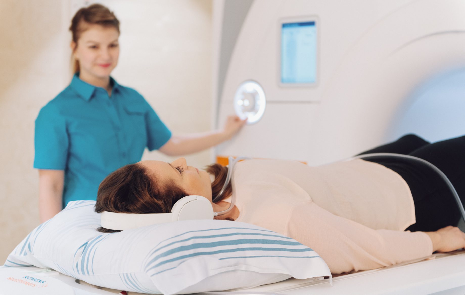
At Docrates, MRI scans are conducted with next-generation MRI scanners. We use the Siemens Magnetom Vida 3T with imaging power of three teslas. The 3 teslas provide more accuracy when diagnosing breast cancer. The better images of the new MRI scanner enable the physicians to make a more detailed diagnosis. This also helps us draw up even better treatment plans.
A breast MRI uses a breast coil which is placed as close to the imaged area as possible. An intravenously administered contrast agent is also used in imaging.
Our skilled and experienced radiologists, breast cancer surgeons and oncologists work in close cooperation under one roof, making the communication between them efficient and immediate.
As a screening method, a breast MRI is particularly suited for young women potentially at risk of hereditary breast cancer, for women with densely structured breasts as well as for situations where mammography is considered unpleasant. The scan does not expose you to radiation, and dense mammary glands do not hinder the detection of changes in the tissue, unlike in mammography.
A temporary technical problem occurred in the data network connections of Docrates Cancer Center. We apologise for any delays caused...
Read more
Hannu Nurmio was aware that prostate cancer is the most common cancer in older men. However, after the diagnosis was confirmed,...

Early detection of colorectal cancer is crucial for successful treatment. If diagnosed early, up to 90% of cancers can be...

Docrates Cancer Center is the first service provider in the Nordic region to launch a new experimental alpha radiation treatment...
Contact us!
Mon-Thu 8:00-18:00, Fri 8:00-16:00
